The Hypothalamus and the Regulated Body
14.1 The adaptive problem: regulating through other bodies
The regulatory systems in the previous chapters defended variables that can be measured inside one body: core temperature, plasma osmolarity, circulating fuel, and stored energy. Social and parenting behavior are different. There is no single social molecule in the blood, no simple equivalent of sodium concentration or leptin. Yet for mammals, social contact and caregiving are not optional decorations added on top of physiology. They are among the ways the body stays regulated.
For a group-living mammal, other animals are part of the survival environment. They provide warmth, protection, information about food and danger, mating opportunity, cooperative defense, and, in many species, help with care. Isolation can therefore become a biological problem. A solitary animal may be colder, more vulnerable to predators, less able to detect danger, less able to share information, and less buffered against stress. In humans, social isolation and loneliness are associated with changes in stress physiology, immune function, sleep, cognition, and health. The important point for this unit is not that social life is emotionally meaningful, although it certainly is. The point is that social contact changes the body’s regulatory situation.
Parenting raises the stakes further. Mammalian offspring are often born unable to feed themselves, defend themselves, or maintain body temperature. Many are altricial: neurologically immature, motorically weak, and dependent on prolonged care. A newborn mammal cannot wait until its own temperature, glucose level, or hydration has failed before help arrives. The parent’s body and behavior must become part of the offspring’s regulatory system. A nursing mother does not simply produce milk; she detects infant cues, releases oxytocin, ejects milk, warms the infant, retrieves it when it wanders, suppresses incompatible behaviors, and stays near enough for the infant’s fragile physiology to remain stable.
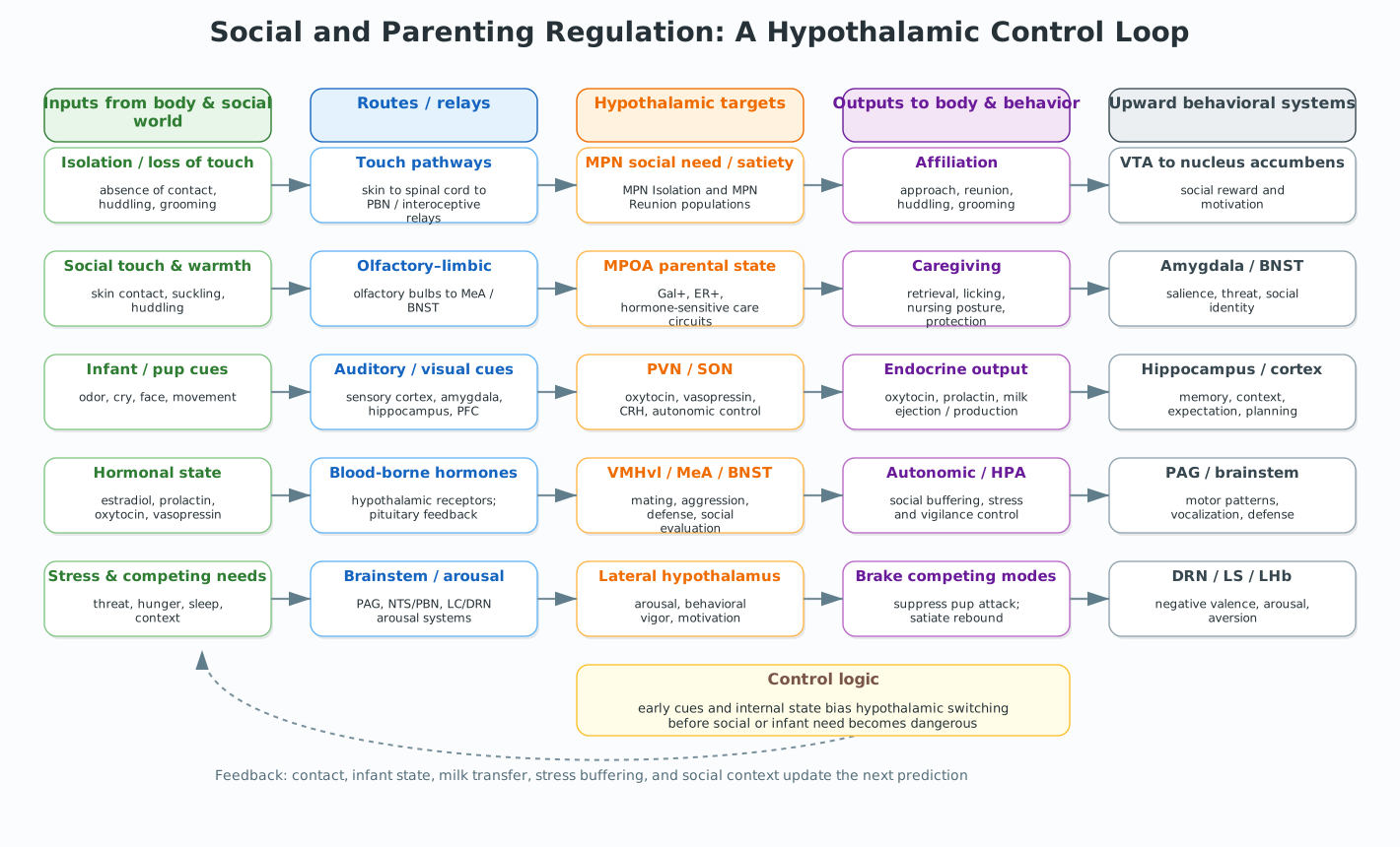
This makes social and parenting behavior a natural, but more speculative, extension of the control-system framework. The adaptive problem is not to hold a single internal variable at a set point. It is to regulate a relational state: enough contact with the right individuals, enough separation when threat or conflict makes contact dangerous, enough caregiving for dependent offspring, and enough flexibility to switch among affiliation, care, mating, aggression, defense, and withdrawal. The same conspecific can be a partner, competitor, infant, intruder, offspring, threat, or source of comfort depending on context and internal state. The hypothalamus is well placed for this problem because it links sensory input, hormonal state, autonomic output, endocrine output, and motivated behavior.
The phrase social homeostasis is useful here, but it must be used carefully. Matthews and Tye proposed that social connection can be treated as an innate need: the brain monitors the quantity and quality of social contact, detects deviation from an expected range, and recruits behavioral and physiological effectors to restore contact. That framework is conceptually strong. It explains why loneliness might feel aversive rather than merely sad: the feeling may function as a drive state, pushing the animal toward reconnection in the way hunger pushes it toward food.
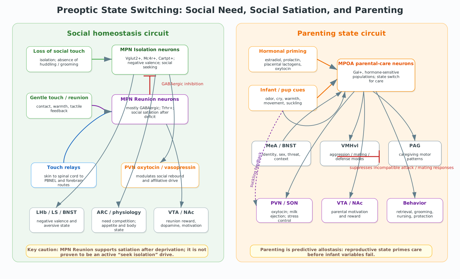
The empirical evidence is strongest for one direction of the loop. Acute social isolation can produce a measurable social-need state. When isolated rodents are reunited with familiar animals, they show a rebound in social interaction, and that rebound gradually satiates as reunion proceeds. Recent mouse work has identified preoptic hypothalamic neurons that are active during isolation, opposing neurons that are active during reunion, and a tactile signal that helps tell the brain that social contact has occurred. That is a major step toward a mechanistic social-homeostasis circuit.
The evidence is weaker for the mirror-image claim that “too much society” activates the same system in reverse and produces an active drive for isolation. Crowding and social overload can certainly produce withdrawal, aggression, stress responses, and avoidance. But those phenomena are not yet clearly the opposite arm of the same social homeostat. They may involve sensory load, dominance relationships, competition, serotonin, stress physiology, and species-specific social ecology. In this chapter, we will therefore treat social homeostasis as best supported for deficit correction: isolation produces need, reunion produces satiation, and chronic or excessive social stress may recruit partly different systems.
In ordinary language, “social satiety” can sound as though the animal has had too much contact and now wants isolation. In the strongest current circuit evidence, however, social satiety has a narrower meaning. It means that a prior deficit has been reduced. After isolation, reunion gradually lowers the rebound in social interaction; the animal no longer behaves as though it has an accumulating social need.
That is analogous to hunger. Eating a meal reduces hunger, but satiety is not automatically an aversive drive to avoid all food. Likewise, activating reunion-associated neurons in the medial preoptic nucleus reduces social rebound after isolation, but does not appear to suppress ordinary social interaction in group-housed animals. That distinction matters. It lets us use the homeostatic framework without overstating the symmetry of the evidence.
14.2 Sensors and signals: cues of isolation, reunion, offspring, and state
Social and parenting circuits have to solve a harder sensing problem than osmolarity or glucose. They must detect not only whether another body is present, but who that body is, what it means, what state it is in, and what the current animal can afford to do about it. The relevant inputs therefore arrive through several channels at once: tactile contact, warmth, smell, sound, vision, hormonal state, prior history, and learned context.
14.2.1 Touch, warmth, and contact
The most direct signal of social contact is physical contact. Huddling, grooming, nursing, carrying, cuddling, and gentle touch all provide information that another body is near enough to matter. This is especially clear in rodents, where much of social behavior occurs through the body surface: sniffing, head-to-head contact, crawling under a cagemate, allogrooming, pup retrieval, and nursing posture.
The recent Liu et al. study makes touch central to social homeostasis in mice. The investigators separated adult female sibling mice and then measured social rebound during reunion. If a perforated divider allowed auditory and olfactory cues but prevented direct contact, mice still showed a robust social rebound when reunited. Impaired vomeronasal pheromone sensing did not abolish rebound or its satiation. By contrast, reducing mechanosensory input delayed the satiation of social need, and genetic manipulations affecting touch-related sensory neurons reduced social rebound. Soft cloth contact could partly substitute for social touch: after isolated mice crossed soft, fur-like tunnels, their later social rebound was reduced, whereas crossings through naked tunnels did not have the same effect.
This does not mean that a single “pleasant touch” fiber runs straight to a hypothalamic social center. The route is distributed. Touch signals begin in peripheral somatosensory afferents, ascend through spinal and brainstem pathways, engage parabrachial and thalamic relays, and interact with hypothalamic, limbic, and reward circuits. But the control logic is clear: for mice, the absence of touch helps signal isolation, and the presence of gentle contact helps signal reunion.
That sensory arrangement also explains why social contact is not equivalent to mere information about another animal. Hearing or smelling a cagemate through a divider says that another animal exists nearby. Touch says something stronger: the other animal is close enough for warmth, grooming, nursing, protection, and affiliation. For a small mammal, that distinction has regulatory consequences.
14.2.2 Identity, familiarity, and infant cues
Social regulation also requires identity. A parent must distinguish offspring from prey or intruders. A pair-bonded animal must distinguish a partner from a stranger. A group-living animal must distinguish familiar cagemates from unfamiliar competitors. These distinctions are not all made in the hypothalamus, but hypothalamic social circuits depend on them.
In rodents, olfactory and vomeronasal cues are especially important for identity, reproductive status, and social category. These signals are processed through olfactory structures, the medial amygdala (MeA), and the bed nucleus of the stria terminalis (BNST), which then communicate with hypothalamic nodes such as the medial preoptic area, ventromedial hypothalamus, and premammillary regions. Pup odors, adult pheromonal cues, and familiar cagemate odors can therefore bias the hypothalamus toward care, mating, aggression, or affiliation depending on internal state.
Humans rely less on vomeronasal signaling and more on visual, auditory, tactile, and learned cues. Infant cries, facial expressions, body posture, skin-to-skin contact, and the learned meaning of a particular child or partner all contribute to parental and affiliative responses. These routes involve cortex, amygdala, hippocampus, striatum, and brainstem as well as hypothalamus. The same principle still applies: the hypothalamus does not respond to a generic social stimulus. It receives a socially interpreted stimulus, already shaped by sensory systems, memory, and context.
Infant cues are especially important because they are predictive. A cry can signal hunger, pain, cold, separation, or distress before the infant’s internal variables have failed. Touch and suckling signal that the infant is present and feeding. Odor and warmth signal proximity. In a primed parent, these cues do not merely produce perception; they trigger a caregiving mode.
14.2.3 Hormonal state and reproductive priming
The meaning of infant cues depends strongly on hormonal state. Pregnancy, birth, lactation, sexual experience, and prior caregiving all change how the brain responds to offspring. Estradiol, progesterone, prolactin, placental lactogens, oxytocin, vasopressin, and stress hormones all influence preoptic and hypothalamic circuits. These signals are slower than touch or sound, but they set the gain of the system.
This is a classic allostatic design. The mother is not primed only after the infant has become cold or hungry. Hormonal changes late in pregnancy and around birth prepare the brain in advance, lowering the threshold for infant-directed care and changing the motivational value of pup or infant cues. Suckling then closes the loop: sensory input from the nipple reaches hypothalamic oxytocin neurons, oxytocin is released from the posterior pituitary, milk is ejected, and infant contact continues.
The paraventricular nucleus (PVN) and supraoptic nucleus (SON) are central to this endocrine arm because they contain magnocellular neurons that produce oxytocin and vasopressin. Oxytocin is released into the bloodstream from the posterior pituitary, where it supports milk ejection and uterine contraction, but oxytocin neurons also project within the brain. Those central projections can modulate social recognition, affiliation, reward, stress, and parental behavior. Vasopressin, closely related to oxytocin, also participates in social recognition, pair bonding, territorial behavior, and paternal behavior in some species.
It is tempting to summarize this by saying that oxytocin is the “bonding hormone.” That slogan is too simple. Oxytocin is not love in molecular form. Its effects depend on receptor distribution, sex, species, social history, stress state, and the circuit in which it acts. In some contexts it promotes approach, recognition, and trust-like behavior; in others it can sharpen social salience or intensify responses to in-group versus out-group cues. In this chapter, oxytocin is best treated as a state-dependent modulator that helps social stimuli matter more.
14.2.4 Context, expectation, and competing needs
Finally, social regulation depends on expectation. A solitary animal in a normally solitary species is not in the same state as a group-living animal separated from its cagemates. A parent temporarily away from an infant is not in the same state as a non-parent encountering a pup. A person alone by choice is not necessarily in the same state as a person who feels excluded. Social need depends on quantity, quality, predictability, identity, and history.
Other physiological needs also change the social set point. Hunger may suppress social exploration and make food the dominant target. Thirst may increase tolerance of social contact at a communal water source. Predatory threat may make grouping more valuable. Infection may reduce social approach to limit energy expenditure or disease spread. Parenting can make offspring cues override sleep, hunger, mating, and self-protection. The social controller is therefore not a separate module bolted onto the rest of homeostasis. It is part of a motivational hierarchy in which competing needs are continuously weighted against each other.
14.3 Hypothalamic circuits: preoptic state switching
The social and parenting circuits are distributed, but the preoptic hypothalamus provides a useful point of entry. This anterior hypothalamic region includes the medial preoptic nucleus (MPN) and medial preoptic area (MPOA), and it participates in thermoregulation, sleep, reproduction, parenting, and social behavior. That overlap is not an accident. The preoptic region sits at a place where bodily state, reproductive hormones, sensory signals, and motivated action converge.
The old “center” language is especially dangerous here. The MPOA is not a parenting center, the PVN is not a bonding center, and the VMHvl is not simply an aggression center. Each is a node in a broader circuit. What makes the preoptic region important is not that it contains a single command button for social behavior. It is that it can switch behavioral state by integrating cues, hormones, and competing drives.
14.3.2 The MPOA parenting circuit
Parenting uses overlapping anatomy but a different regulatory problem. The parent is not simply correcting its own social deficit. It is entering a state in which offspring cues become urgent, rewarding, and action-commanding. The medial preoptic area is the best-studied hypothalamic node in this transition.
In many mammals, pup or infant cues do not have a fixed meaning. In one state, a pup may be ignored. In another, it may evoke avoidance or even attack. In a hormonally and experientially primed parent, the same cues evoke approach, retrieval, licking, grooming, nursing posture, nest building, and protection. This state switch depends on the MPOA, but not because the MPOA stores a complete parenting program. Rather, it integrates reproductive hormones, infant sensory cues, stress state, prior experience, and competing social circuits.
Modern causal work has identified MPOA galanin neurons as a key parental-care population in mice. These neurons are activated by pup exposure in parents and participate in both maternal and paternal care. Loss of function disrupts parental behavior, while activation biases animals toward pup-directed care and away from pup-directed aggression. This is a powerful example of the control logic emphasized throughout this unit: the same stimulus can lead to opposite behavior depending on hypothalamic state. A pup is not simply “recognized.” It is assigned a biological meaning.
MPOA outputs are distributed. Projections to PAG and other midbrain/brainstem regions help organize motor patterns such as crouching, grooming, retrieval, and defensive protection. Projections to VTA and nucleus accumbens help make pup cues motivationally valuable, so the animal will approach, retrieve, and work for contact. Connections with PVN and SON link caregiving to oxytocin release, lactation, milk ejection, and social buffering. Connections with amygdala, BNST, and VMHvl help suppress incompatible responses such as attack, mating, or flight when infant care should dominate.
Again, the important word is state. Parenting is not a reflex released by a pup odor. It is a whole-body regulatory mode. Hormones prepare the system in advance. Infant cues trigger and maintain it. Suckling and touch feed back to the hypothalamus. Reward circuits make caregiving persistent. Stress circuits are modulated so that infant distress is not treated merely as a threat. The behavior is social, but the architecture is regulatory.
14.3.3 Opponent circuits for care, mating, aggression, and defense
The hypothalamus must also prevent incompatible social programs from running at the same time. A mouse cannot retrieve a pup and attack it simultaneously. A parent cannot devote the same motor system to nursing, mating, and fleeing. Social regulation therefore depends heavily on inhibition and disinhibition.
The ventrolateral ventromedial hypothalamus (VMHvl) participates in aggression, mating, and social defensive behaviors. The medial amygdala and BNST provide information about sex, reproductive status, familiarity, threat, and social category. These regions are not “bad” circuits opposed to a “good” parenting circuit. They are essential for adaptive social behavior. But during parenting, their outputs must be gated so that pup-directed care can dominate over pup-directed attack or mating-related behavior.
This opponent logic is familiar from earlier chapters. AgRP neurons promote hunger partly by inhibiting satiety pathways. Thirst circuits are suppressed by drinking-related feedback. Sleep and wake circuits inhibit each other to stabilize state transitions. Parenting and social homeostasis use a similar principle: the brain does not merely activate the desired behavior. It also suppresses competing states that would make that behavior impossible.
14.4 The affective and behavioral drive
A regulatory state becomes behavior by changing what the world means. Hunger makes food cues more salient. Thirst makes water cues compelling. Social need makes contact valuable, and parenting makes infant cues urgent. This is the affective side of hypothalamic regulation: the body state is translated into a motivated state.
Acute isolation illustrates the point. The isolated animal is not simply missing a social stimulus. It enters a state with negative valence, heightened attention to social opportunity, and increased willingness to interact when reunion becomes possible. Matthews and Tye organized this response into three overlapping components: hypervigilance or arousal, social motivation, and passive coping. Those are not three unrelated effects. They are three ways a social deficit can reorganize the animal. Being alone can make the environment feel more dangerous, make social contact more valuable, and, if reconnection fails, shift the animal toward coping strategies that limit distress.
The Liu et al. circuit gives this framework a hypothalamic anchor. MPNIsolation activity carries negative valence and promotes social interaction. MPNReunion activity carries positive valence and helps satiate the need. VTA and nucleus accumbens dopamine systems participate in the rewarding and motivational aspects of reunion. Dorsal raphe dopamine neurons, discussed by Matthews and Tye, may contribute to the aversive drive of isolation: stimulating them can increase social preference when a social target is available, yet be avoided when no social stimulus is present. That is a useful distinction for students. Social seeking after isolation is not simply pleasure seeking. It may also be relief seeking.
Parenting has a parallel motivational transformation. In a non-parent, an infant’s cry may be irritating, alarming, or irrelevant. In a primed caregiver, the same cry becomes action-guiding. Pup odors, infant faces, suckling, and touch acquire motivational significance. The parent is willing to interrupt sleep, feeding, mating, and exploration to retrieve, warm, feed, and protect the offspring. The reward system is not outside the regulatory loop; it is one way the loop gives priority to care.
This state dependence is a form of social alliesthesia. A stimulus is not rewarding or aversive in isolation from the body. Its value depends on internal state. A pup is not the same stimulus to a virgin male mouse, a lactating mother, and a father with caregiving experience. A familiar cagemate is not the same stimulus to a recently isolated mouse and a mouse that has been group-housed. A partner is not the same stimulus in safety, threat, hunger, illness, or reproductive readiness. The hypothalamus helps set those meanings by controlling the internal state in which the stimulus is encountered.
Social reward also depends on memory and specificity. In pair-bonding species such as prairie voles, oxytocin, vasopressin, dopamine, and nucleus accumbens circuits help attach reward to a particular partner rather than to any conspecific. In humans, social value is even more strongly shaped by memory, culture, language, and expectation. But the deep logic remains biological: social stimuli become motivationally powerful because they predict regulation, protection, mating, offspring survival, and belonging.
14.6 Clinical failures and human relevance
Clinical examples are useful here because they reveal the architecture, but they must be handled cautiously. Social and parenting behavior are not controlled by a single hypothalamic nucleus, and human disorders are not simple failures of one mouse circuit. The more useful question is what part of the control loop has become unstable: sensing, valuation, hormonal priming, stress regulation, reward, memory, or behavioral output.
Loneliness and chronic isolation are the clearest human relevance of social homeostasis. Acute isolation may produce an adaptive drive to reconnect. Chronic isolation is different. When reconnection fails, the same systems that promote vigilance, stress readiness, and social motivation may remain engaged too long. That can contribute to sleep disruption, HPA-axis dysregulation, inflammatory changes, anxiety, depressive symptoms, and cognitive strain. The distinction between acute and chronic isolation matters because a drive state that is adaptive over hours or days can become damaging when sustained for months or years.
Postpartum depression and related peripartum disorders show how parenting depends on a distributed state transition. A simple explanation would be that the MPOA fails to turn on. That is too narrow. Postpartum depression involves interactions among hormonal change, sleep disruption, stress, prior vulnerability, reward processing, social support, and the sensory salience of infant cues. Infant cries may become overwhelming rather than action-guiding; caregiving may feel effortful rather than rewarding; stress physiology may not be adequately buffered by contact. The hypothalamus is likely part of this architecture, especially through stress, oxytocin, prolactin, sleep, and reproductive-hormone pathways, but it is not the whole explanation.
Autism spectrum conditions are also relevant but easy to oversimplify. Oxytocin, vasopressin, dopamine, and social reward have all been studied in relation to social motivation and social salience. But autism is heterogeneous, developmentally complex, and not reducible to a broken “bonding hormone” system. For some individuals, social stimuli may be less rewarding; for others, they may be overwhelming, unpredictable, or difficult to interpret. The control-system language can help only if it respects that diversity: the problem may lie in sensory prediction, salience, social learning, stress, motor timing, communication, or combinations of these, not in one hypothalamic social switch.
Early adversity and neglect show how social regulation is built through repeated feedback. Touch, warmth, predictable caregiving, feeding, and stress buffering shape the developing child’s expectation that other bodies can regulate distress. Severe neglect can disrupt stress regulation, attachment behavior, sleep, feeding, and later social trust. It is reasonable to think about this as a disturbance in developing social allostasis: the infant’s brain is learning what social cues predict and whether contact restores safety. But claims about permanent damage to a single receptor system should be avoided. Development remains plastic, and interventions work partly by restoring reliable regulatory relationships.
Hypothalamic or pituitary injury can also disturb parenting and social physiology indirectly. Damage that alters sleep, thermoregulation, stress hormones, gonadal hormones, prolactin, oxytocin, appetite, or autonomic regulation can change social behavior even if social perception remains intact. This reinforces the main lesson of the unit: motivated behavior is built on bodily regulation. When the body-control systems fail, the social world changes too.