13 Reproduction
The Hypothalamus and the Regulated Body
13.1 The adaptive problem: timing an expensive investment
Reproduction is not a single internal variable like temperature, osmolarity, or blood glucose. There is no one number that the body holds near a set point called “reproduction.” The regulated object is a state: a coordinated condition in which the body is mature enough, energetically able, hormonally prepared, socially cued, and behaviorally motivated to invest in the next generation.
That investment is expensive. Gametes must be produced. Steroid hormones must organize tissues and alter behavior. In females of many species, ovulation, pregnancy, and lactation impose enormous energetic costs. In males, spermatogenesis, testosterone-dependent physiology, courtship, mate competition, and risk-taking also carry costs. In both sexes, sexual behavior can expose an animal to predation, injury, disease, and social conflict. Reproduction is therefore not simply a behavior added on top of survival. It is a biological allocation decision: how much of the body’s limited budget should be spent now on future offspring rather than on immediate maintenance?
This makes reproduction an especially clear example of allostasis. A purely reactive controller would be useless. Puberty must be prepared years before adult reproduction. Ovulation must be timed before conception can occur. Gonadal tissues must be maintained before a mating opportunity appears. Sexual motivation must be biased by social cues before copulation begins. The body cannot wait until the full cost of reproduction is already present before deciding whether it can afford that cost.
The hypothalamus sits at the center of this timing problem. It does not act as a simple “sex center.” It integrates energy reserve, stress, circadian and seasonal timing, gonadal steroid feedback, sensory cues from potential partners, and learned social context. It then adjusts endocrine output through the hypothalamic-pituitary-gonadal axis, autonomic output to the reproductive organs, and behavioral output through motivational and motor systems. The result is not a reflex but a state switch. Under some conditions the reproductive axis is permitted and sexual stimuli become salient. Under other conditions the same axis is suppressed, and reproduction is delayed in favor of survival.
The deepest link is with energy balance. In the previous chapter, we treated energy as the budget from which other regulatory systems spend. Reproduction is one of the most expensive purchases made from that budget. Low energy reserve can delay puberty, suppress ovulation, lower gonadal steroid output, reduce sexual motivation, and narrow behavior toward food seeking. This is not merely pathology. It is the normal logic of a controller that asks whether the body can afford to reproduce.
13.2 Sensors and signals: internal permission and external opportunity
Reproductive control depends on several streams of information that operate on different timescales. Some signals report whether the body is mature and energetically prepared. Others report whether the gonads are functioning and where the animal is in a cycle. Still others report whether a possible mate is present, safe, and appropriate. The hypothalamus has to combine all of them because none is sufficient alone.
13.2.1 Metabolic and stress signals
The first question is whether reproduction is biologically affordable. Leptin, secreted by adipose tissue, is a major permissive signal. It does not simply “turn on” reproduction by itself, and GnRH neurons are not just leptin meters. But low leptin is a strong warning that fat reserve is inadequate. In that state, the reproductive axis is often suppressed. Puberty may be delayed, menstrual or estrous cycling may stop, and gonadal steroid levels may fall. This is the same conservation program introduced in the energy-balance chapter: when reserve is low, the body spends less on growth and reproduction.
Other metabolic signals contribute to the same estimate. Insulin, ghrelin, thyroid status, nutrients, and the general pattern of energy availability all influence reproductive function. These signals reach the hypothalamus through blood-borne routes, local nutrient-sensitive neurons, and brainstem-visceral pathways. Their control logic is permissive rather than commanding. Adequate energy does not guarantee reproduction, but inadequate energy can veto it.
Stress provides a second veto. The hypothalamic-pituitary-adrenal axis and the reproductive axis are not independent systems running in parallel. Stress-related signals, including CRH, cortisol, endogenous opioids, and inflammatory signals, can suppress pulsatile GnRH release and reduce gonadotropin output. That tradeoff is adaptive in principle. Severe danger, illness, famine, or chronic social threat are poor times to invest in reproduction. But the same rule can become maladaptive in modern humans when chronic psychological stress, excessive exercise, or undernutrition suppresses reproductive function even in the absence of immediate danger.
13.2.2 Gonadal steroid feedback
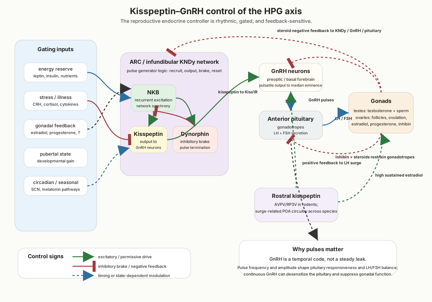
The reproductive axis also monitors its own downstream products. The gonads produce sex steroids: estradiol, progesterone, and testosterone. These hormones act throughout the body, but they also feed back to the hypothalamus and pituitary. In the simplest negative-feedback mode, rising steroid levels restrain further hypothalamic and pituitary drive. The brain asks, in effect, whether the gonads are already producing enough hormonal output for the current state.
In females of cycling species, estradiol also has a more unusual role. For much of the cycle, estradiol participates in negative feedback. But when estradiol remains high in the appropriate preovulatory context, the system reverses sign: sustained estradiol produces positive feedback that drives a surge of GnRH and luteinizing hormone. That surge is what triggers ovulation. This is a beautiful control problem because the same hormone can mean different things depending on timing, concentration, and circuit state. Estradiol is not simply an “on” or “off” signal. It is interpreted by hypothalamic networks that know where the body is in the reproductive cycle.
Sex steroids also act on hypothalamic and limbic circuits that regulate behavior. Testosterone, estradiol, and progesterone alter the sensitivity of the medial preoptic area, ventromedial hypothalamus, medial amygdala, BNST, and downstream motor systems. Some effects are organizational, shaping circuits during development. Others are activational, changing adult motivation and receptivity over hours, days, or weeks. Keeping these two timescales separate prevents a common mistake: adult hormones modulate reproductive behavior, but they act on circuits whose structure was partly organized earlier in life.
13.3 Hypothalamic circuits: opening and closing the reproductive axis
The reproductive system has two linked circuit layers. The first is endocrine: hypothalamus to pituitary to gonads and back again. The second is behavioral: hypothalamic and limbic circuits that alter sexual motivation, social investigation, receptivity, autonomic arousal, and motor patterning. These layers are separable for teaching, but in the living animal they are coupled. Hormonal state changes behavior, and behavioral context changes hormonal output.
13.3.1 GnRH neurons and the HPG cascade
The endocrine layer begins with a small population of gonadotropin-releasing hormone neurons. These neurons are unusual in both development and function. During embryonic life, GnRH neurons arise outside the brain in the nasal/olfactory region and migrate into the forebrain and hypothalamus. In adulthood, their axons release GnRH into the capillary bed of the median eminence, a specialized interface where hypothalamic signals enter the hypophyseal portal circulation. GnRH then travels a short distance to the anterior pituitary.
The anterior pituitary responds by releasing the gonadotropins luteinizing hormone and follicle-stimulating hormone. LH and FSH act on the gonads. In the testes, LH stimulates Leydig cells to produce testosterone, while FSH acts on Sertoli cells and supports spermatogenesis and inhibin production. In the ovaries, FSH supports follicular growth and estradiol production, while LH contributes to steroidogenesis and, at the right moment, triggers ovulation and luteal function. The gonads then send feedback to the brain and pituitary through sex steroids and inhibin.
A crucial feature of this axis is pulsatility. GnRH is not simply released at a constant level. It is released in pulses, and the frequency and amplitude of those pulses matter. Pulsatile GnRH maintains pituitary responsiveness. Continuous GnRH stimulation, paradoxically, can desensitize pituitary gonadotropes and suppress LH and FSH release. Clinicians exploit this property with long-acting GnRH agonists in several reproductive and hormone-sensitive conditions. For the textbook point, pulsatility shows that reproduction is controlled by temporal pattern, not just hormone concentration.
13.3.2 Kisspeptin, KNDy neurons, and pulse control
GnRH neurons are the final hypothalamic output to the pituitary, but much of the control sits upstream. The most important upstream signal is kisspeptin, acting through the kisspeptin receptor on GnRH neurons. Loss-of-function mutations in kisspeptin signaling can produce failure of puberty and hypogonadotropic hypogonadism, while increased kisspeptin drive is associated with activation of the reproductive axis.
A major source of this drive is the arcuate nucleus in rodents and the homologous infundibular region in humans. Many of these neurons co-express three peptides: kisspeptin, neurokinin B, and dynorphin. They are often called KNDy neurons. The three-peptide arrangement gives the circuit a natural oscillator. Neurokinin B provides excitatory synchronization among KNDy neurons. Dynorphin supplies an inhibitory brake that helps terminate each burst. Kisspeptin is the output that stimulates GnRH neurons. The result is rhythmic GnRH drive, which the pituitary reads as pulsatile LH and FSH release.
This is the reproductive counterpart of the melanocortin circuit in energy balance. The important lesson is not the memorization of peptide names. It is the logic: a hypothalamic population integrates metabolic and steroid feedback, uses local excitation and inhibition to generate pulses, and drives an endocrine cascade only when the body state permits it.
13.3.3 Positive feedback and the ovulatory surge
The same kisspeptin-GnRH system also helps explain why ovulation requires a switch in feedback sign. In cycling females, estradiol from the growing follicle initially restrains gonadotropin drive. But sustained high estradiol in the appropriate phase activates a positive-feedback pathway that produces the preovulatory GnRH/LH surge. In rodents, a rostral hypothalamic kisspeptin population in the anteroventral periventricular and periventricular regions is especially important for this surge. In humans, the anatomy is not identical, and infundibular KNDy neurons appear to participate in both negative and positive feedback. The species difference matters, but the control principle is shared: the reproductive axis can reverse from restraint to amplification when the hormonal pattern predicts imminent ovulation.
That reversal makes sense only if the system is read as predictive control. Ovulation is not a correction for an error. It is a timed event. The brain and pituitary must coordinate a future possibility: release an oocyte when follicular development, steroid state, and behavioral conditions are favorable. The LH surge is therefore an endocrine decision point, not a passive consequence of high estradiol alone.
13.3.4 MPOA, VMHvl, MeA, BNST, and reproductive behavior
The behavioral layer of reproductive control is distributed across hypothalamic, amygdalar, striatal, midbrain, and spinal circuits. The medial preoptic area is one of the major hypothalamic nodes. In male rodents, MPOA lesions severely impair copulatory behavior, and MPOA stimulation or hormonal priming can facilitate sexual behavior. The MPOA receives steroid, olfactory, somatosensory, and motivational input, and it projects to the VTA, periaqueductal gray, brainstem autonomic nuclei, and spinal systems. It is therefore better understood as an integrative reproductive-action node than as a male “sex center.”
The ventrolateral ventromedial hypothalamus is another major node, especially in female sexual receptivity in rodents. Estradiol primes VMHvl circuits in part by inducing progesterone receptor expression, and progesterone can then help open the behavioral window for lordosis, the reflexive posture that permits copulation in many quadrupeds. VMHvl lesions or disruption of estrogen receptor signaling impair this receptive behavior. But the VMHvl is not simply a female receptivity center. Neighboring and partly overlapping VMHvl populations also participate in aggression, social investigation, and defensive decisions. The same region helps decide whether another animal is approached, accepted, mounted, attacked, or avoided.
The medial amygdala and bed nucleus of the stria terminalis are essential relays for social information. In rodents, chemosensory information about sex, reproductive status, and individual identity reaches the MeA and BNST before influencing MPOA and VMHvl. These structures help convert sensory evidence about another animal into hypothalamic action tendencies. In humans, the sensory routes are more visual, auditory, tactile, olfactory, and learned, but the larger principle remains: reproductive motivation is embedded in social perception and social memory.
Modern causal studies have sharpened this picture. Optogenetic and chemogenetic experiments in mice show that molecularly defined hypothalamic neurons can bias specific components of reproductive and social behavior. For example, Esr1-expressing VMHvl neurons are active during social investigation, mounting, and aggression-related behaviors, and manipulating them can shift the probability or intensity of those behaviors depending on sex, context, and stimulation pattern. Other work has identified preoptic and BNST populations that influence male mating, sexual reward, and partner-directed approach. These experiments are powerful because they do not merely label anatomy. They show how hypothalamic state can change the animal’s behavioral policy toward a social stimulus.
Older textbooks often described reproductive behavior in center language: the MPOA as a male copulatory center, the VMH as a female receptivity center, and the hypothalamus as the command structure for sex. Those labels captured real lesion effects, but they were too simple.
A lesion does not remove only one behavior. It removes a node from a loop. Damage to the MPOA can disrupt sexual motivation, integration of steroid signals, genital feedback, dopamine-dependent approach, autonomic coordination, and projections to the PAG and spinal cord. Damage to the VMHvl can disrupt receptivity, rejection, aggression, defensive behavior, and state-dependent interpretation of social stimuli. The behavioral change is therefore not evidence for a single-purpose center. It is evidence that these hypothalamic regions participate in distributed reproductive-control loops.
The modern view is more useful for this unit. Reproduction requires endocrine permission, sensory identification of a potential partner, affective valuation, autonomic arousal, motor patterning, and feedback from the body. No single nucleus can perform all of those jobs. The hypothalamus contributes the body-state and action-selection core, but it works with the amygdala, BNST, VTA, nucleus accumbens, hippocampus, cortex, PAG, pituitary, gonads, and spinal cord.
13.4 The Affective and Behavioral Drive
Reproductive physiology matters because it changes what the animal is motivated to do. A gonadally mature animal with adequate energy reserve does not merely have different hormone levels. It may investigate social cues more strongly, approach potential partners, tolerate closer contact, display courtship behavior, compete with rivals, or become receptive to stimulation that would otherwise be irrelevant or aversive.
This is another form of alliesthesia. The value of a stimulus depends on the internal state of the body. A smell, touch, vocalization, face, body posture, or context can be attractive, neutral, threatening, or disgusting depending on hormonal state, stress, prior experience, social relationship, and learned meaning. The sensory stimulus is not evaluated in isolation. It is evaluated by a nervous system whose hypothalamic and endocrine state has changed the significance of the stimulus.
The MPOA and VMHvl connect reproductive body state to broader motivational systems. Projections to the VTA and nucleus accumbens help assign incentive value to partners, sexual cues, and contexts. Connections with the amygdala and BNST link reproductive motivation to social recognition, threat assessment, and sustained arousal. The hippocampus and cortex contribute memory and context: where a partner was encountered, whether the setting is safe, what social rules apply, and what prior experience predicts. The periaqueductal gray and spinal cord help convert this motivational state into organized action.
This connection to reward should not be misunderstood. Sexual behavior is often reinforcing, and dopamine systems are important. But reproductive motivation is not reducible to reward in the abstract. It is a body-state system coupled to reward and social cognition. The hypothalamus biases the animal toward reproductive action when internal permission and external opportunity align. Telencephalic systems then help determine the object, context, cost, and learned value of that action.
This is also why reproductive motivation can be suppressed. Low energy reserve, severe stress, illness, pain, fear, social defeat, or unsafe context can reduce sexual interest even when the gonads are capable of producing hormones. The reproductive system is not trying to maximize mating at all times. It is trying to time mating so that the costs are worth paying.
Parenting and caregiving are related but not identical regulatory problems. They require their own hypothalamic and social-control loops, including strong contributions from preoptic, hormonal, sensory, and reward systems. This chapter focuses on reproductive readiness and sexual behavior; the next chapter takes up social and parenting regulation more directly.
13.5 Effectors: endocrine, autonomic, motor, and behavioral
The outputs of reproductive control are broad because reproduction is not solved by one effector. The hypothalamus must regulate endocrine state, gonadal tissue, genital blood flow, pelvic reflexes, courtship, social approach, and sometimes suppression. These outputs unfold over timescales from seconds to years.
The endocrine arm is the HPG axis. GnRH pulses control pituitary LH and FSH. LH and FSH regulate the gonads. The gonads produce gametes and sex steroids. Steroids then act on the body to support secondary sexual characteristics, reproductive tissue function, bone and muscle physiology, libido, and feedback to the brain and pituitary. In ovaries, the cycle requires coordinated follicular development, ovulation, luteal function, and progesterone production. In testes, continuous spermatogenesis depends on local testosterone, Sertoli cell function, and FSH support. Inhibin provides additional feedback, especially on FSH.
The autonomic arm adjusts the reproductive organs. Parasympathetic pathways support erection, clitoral engorgement, vaginal lubrication, and genital vasocongestion. Sympathetic pathways contribute to emission and ejaculation in males and to several genital and uterine responses in females. These autonomic effects are not separate from motivation. They prepare peripheral tissue for behavior and provide sensory feedback that can sustain arousal.
The somatic and motor arm organizes action. In rodents this includes mounting, intromission, pelvic thrusting, ejaculation, lordosis, rejection, and approach or avoidance. In humans the motor patterns are more flexible and strongly shaped by cognition, relationship, consent, culture, and learning, but the basic point remains: hypothalamic reproductive state must be coupled to motor systems. The PAG, brainstem, and spinal cord are central parts of that coupling.
The behavioral arm changes the animal’s priorities. Reproductive state can increase social investigation, mate seeking, courtship, risk tolerance, competition, and attention to sexually relevant cues. It can also produce rejection or avoidance when conditions are wrong. From the controller’s perspective, both approach and suppression are effectors. A system that cannot inhibit reproduction when the body is depleted or the context is dangerous is as poorly regulated as a system that cannot initiate reproduction when conditions are favorable.
Every reproductive effector has tradeoffs. Sex steroids alter muscle, bone, cardiovascular physiology, immune function, and behavior. Mating can compete with feeding, sleep, thermoregulation, predator avoidance, and social stability. Pregnancy and lactation, where they occur, impose even greater energetic demands. The hypothalamus therefore regulates reproduction not as an isolated drive but as one claim among many on the body’s resources.
13.6 Clinical failures and human relevance
Clinical disorders reveal the architecture by breaking different links in the loop.
Functional hypothalamic amenorrhea is one of the clearest examples. Menstrual cycling can stop when energy availability is too low, exercise load is too high, psychological stress is severe, or some combination of these conditions persists. The proximate mechanism is reduced pulsatile GnRH drive and altered LH/FSH secretion. The control logic is easy to see: the brain reads the body as unable to afford reproduction and suppresses the axis. The result can be adaptive in famine but harmful when the trigger is chronic stress, disordered eating, or excessive training in an otherwise food-rich environment.
Kallmann syndrome and related forms of congenital hypogonadotropic hypogonadism break the developmental construction of the axis. GnRH neurons normally arise in the nasal region and migrate into the brain. When that migration or the associated olfactory development fails, puberty may not occur without treatment because the hypothalamus cannot provide adequate GnRH drive to the pituitary. The association with anosmia or hyposmia makes the developmental link visible: the reproductive endocrine system and olfactory system are built together early in embryonic life.
Hyperprolactinemia shows that endocrine state can suppress reproduction from outside the gonadal steroid loop. Elevated prolactin, often from a prolactin-secreting pituitary adenoma or medication effect, can reduce GnRH and gonadotropin secretion. The clinical result may include amenorrhea, infertility, low libido, or hypogonadism. This is not simply a pituitary problem. It shows how one endocrine condition can close the hypothalamic gate on another endocrine axis.
Polycystic ovary syndrome is more complex and should not be reduced to one hypothalamic cause. It commonly involves hyperandrogenism, ovulatory dysfunction, metabolic factors, and altered gonadotropin dynamics, often with relatively increased LH pulsatility. It is useful here because it shows that reproductive control depends on the timing and pattern of endocrine signals, not only their average levels. A change in pulse dynamics can alter ovarian function and feedback, producing a self-reinforcing disorder of the reproductive axis.
Finally, human sexual motivation and orientation remind us to be cautious when translating animal circuit work. Hypothalamic and limbic circuits are surely involved in human sexuality, but human sexual behavior is shaped by cognition, identity, culture, relationship, memory, consent, and conscious experience to a degree that simple rodent circuit diagrams cannot capture. The human hypothalamus is part of the system, not an explanation by itself.
One reason reproduction has long attracted attention in neuroscience is that some hypothalamic regions are sexually dimorphic. The sexually dimorphic nucleus of the preoptic area is larger in male than female rats, and related sex differences have been described in other mammals. In humans, the interstitial nuclei of the anterior hypothalamus have been studied as possible homologous or analogous structures.
The most famous example is INAH-3. Simon LeVay reported in 1991 that INAH-3 volume differed across groups of men and women and was, in that sample, smaller in homosexual men than in heterosexual men. The finding became widely discussed because it seemed to link hypothalamic structure to human sexual orientation.
The interpretation must remain cautious. The study was post-mortem and correlational. Sample sizes were limited. Illness, age, hormone history, and other variables are difficult to control in such work. Later research supports the broader claim that some anterior hypothalamic structures are sexually dimorphic, but no hypothalamic measurement can predict an individual’s sexual orientation. The useful teaching point is narrower: developmental organization of hypothalamic circuits can differ between groups, but complex human sexuality cannot be reduced to the size of one nucleus.
13.7 Integration: reproduction as predictive control
Reproduction shows the hypothalamus acting less like a thermostat and more like a biological allocator. The system does not defend a single variable. It decides whether the body should enter, maintain, or suppress a costly reproductive state.
The logic is allostatic from beginning to end. Developmental signals help time puberty before reproduction is possible. Metabolic signals estimate whether the body can afford reproductive investment. Stress and illness signals can close the gate when immediate survival is threatened. Gonadal steroids provide feedback about the state of the reproductive organs. Kisspeptin and KNDy neurons translate that feedback into pulsatile GnRH drive. Social, sensory, and contextual cues help determine whether a reproductive opportunity is present. MPOA, VMHvl, MeA, BNST, VTA, nucleus accumbens, PAG, spinal cord, and cortex convert bodily readiness into motivated action or active suppression.
Waiting for error would not solve this problem. By the time pregnancy, energetic depletion, social danger, or failed timing has produced a clear internal error, the biological cost has already been paid. The reproductive system therefore relies on early cues and predictive gates. It opens the HPG axis when conditions suggest that reproduction is worth attempting, and it closes the axis when the body or environment makes that investment too risky.
This is why reproduction belongs in a unit on the regulated body. It is not merely about sex hormones or mating behavior. It is about the hypothalamus coordinating the body’s future: when to grow, when to cycle, when to seek a mate, when to inhibit that seeking, and when to conserve resources for survival instead. Reproduction is the moment when the regulated body asks whether it can afford to spend itself on the next generation.
13.2.3 Social, sensory, and contextual cues
Reproduction also depends on external opportunity. In many mammals, social chemical cues are especially important. The vomeronasal organ and the main olfactory system detect information from conspecifics and route it through the accessory olfactory bulb, medial amygdala, BNST, and hypothalamus. These pathways can influence sexual investigation, partner recognition, territorial behavior, and reproductive timing. Rodents are the standard experimental model, but the broader principle applies across many mammals: reproductive circuits read the social environment.
Humans require a cautious note. Human sexual motivation is strongly influenced by sensory, social, emotional, and learned cues, but claims about a functional human vomeronasal organ or simple human pheromones should be treated carefully. The human system relies heavily on ordinary olfaction, vision, touch, memory, culture, and social meaning. The important point for this chapter is not that all species use the same sensory channel. It is that reproductive control combines internal readiness with external opportunity.
Tactile and genital signals provide another stream of feedback. Genital stimulation activates spinal afferents, including pudendal and pelvic pathways, and engages spinal autonomic and motor reflexes. These signals help sustain arousal, coordinate genital blood flow, and organize the motor sequence of copulation. They also interact with hypothalamic and midbrain circuits, especially the medial preoptic area and periaqueductal gray. Sexual behavior is therefore both descending and ascending: hypothalamic state biases the body toward action, and sensory feedback from the body helps maintain and shape that action.
Timing cues matter as well. In seasonal breeders, day length is converted into a hormonal signal through the retina, suprachiasmatic nucleus, and pineal melatonin. This allows the reproductive axis to anticipate whether offspring would be born into a favorable season. Humans are not seasonal breeders in the same strong sense, but circadian timing, sleep, light exposure, and endocrine rhythms still influence reproductive physiology. Again the principle is allostatic: reproductive control uses cues that predict future conditions, not only feedback from the gonads.